MICROPROCESSOR DEVICE FOR DETECTING BLEEDING IN THE FLUSHING SYSTEMS OF UROLOGICAL PATIENTS
1Sevastopol State University
Abstract
The article describes the development and addition of a system for remote monitoring of the functioning of medical droppers and tracking the procedure for removing fluid. The system consists of optical sensors for droplets, fluid elimination and determination of the presence of blood in the liquid being discharged, a microcontroller, a module for receiving and transmitting data via a wireless channel, a central monitor and a smartphone. Various options for the implementation of the system are considered, studies are conducted to design the most effective and error-free sensor for determining the presence of blood.
Keywords: LED emitter, lithotripsy, microcontroller, remote access, sensor, the presence of blood, transparency, turbidity, web server, web-interface, WiFi module
Category: 05.00.00 Technical sciences
Article reference:
Shamkov A.S., Chernega V.S. Microprocessor device for detecting bleeding in the flushing systems of urological patients // Modern scientific researches and innovations. 2021. № 3 [Electronic journal]. URL: https://web.snauka.ru/en/issues/2021/03/94869
 View this article in Russian
 Nowadays, kidney problems are quite common in people. In particular, urolithiasis is a common disease. To extract stones, various methods are used that allow you to crush the stones into small elements, and then, by washing with a special liquid, remove the remains naturally. Thus, after crushing, the patient is injected with a solution intravenously, a special tube is inserted into the patient’s urethra, which ensures the withdrawal of fluid into a certain container. However, this procedure requires constant monitoring by medical personnel. The point is that it is impossible to break a stone into very small pieces, and in some cases they remain large enough to be removed. These stones damage the urethra, causing bleeding. Moreover, they prevent the further excretion of fluid, which requires a doctor’s surgical intervention. Since it is required to constantly monitor the process of fluid elimination, medical personnel cannot perform other functional duties.
In order to increase the efficiency of the work of medical staff, it was decided to create a microprocessor-based device for detecting bleeding in the flushing systems of urological patients. It is planned to add this device to the previously developed medical dropper control system [1]. The device assumes the presence of two sensors: a liquid transparency sensor (for detecting blood), and a liquid removal sensor (for tracking the process of liquid removal and its volume). The device continuously polls the sensors, forming a data packet, which is sent to the computer of the central nurse’s post by a WiFi connection. On this computer, using the web interface, the parameters of the procedure for all patients in the department are displayed. At the same time, as soon as a critical situation is detected (blood in the fluid being withdrawn or stopping fluid excretion), the information is immediately transmitted via a wireless channel to the server (central nurse’s post). Moreover, the same information is duplicated on the smartphones of medical staff. In this way, the timely notification of the medical workers can be ensured.
The designed system consists of a liquid transparency sensor, a liquid withdrawal control sensor, a microcontroller, a WiFi module for receiving data transmission via a wireless channel, an access point that relays data, a central monitor of a nurse’s post and, directly, a smartphone or tablet, which is constantly at the medical staff …
The liquid withdrawal control sensor is built on the principle of the incoming liquid control sensor described in [1]. It performs the same functions, consists of the same elements, and is also connected to the microcontroller in the same way.
The liquid transparency sensor assumes the presence of an emitting LED and a phototransistor. In this case, the study of the choice of the color of the emitter is carried out, as well as the determination of the degree of influence of external factors (illumination) on the operation of the sensor, depending on the color of the emitter.
In the course of development, a study was conducted on methods for detecting the presence of blood in the excreted fluid. Thus, in [2], a chemical and optical method for determining turbidity is described. Based on the analysis of the turbidity of the liquid, it can be concluded that there is blood in the excreted liquid. However, a study of the parameters obtained from the sensor should be carried out when staining the liquid in different colors. The chemical method is good for its accuracy, but it has significant drawbacks: time and complexity. In order to chemically determine the presence of blood in the excreted fluid, it is necessary to take a sample, prepare the necessary solution to ensure a chemical reaction, and, directly, conduct a study. Due to the complexity of this method, it was decided to use an optical one. In [2], modern devices are described for measuring the degree of transparency of a liquid. They have found applications in various spheres of society, such as: determining the degree of transparency of wastewater, sewers, water pipes, rivers, seas and lakes. However, there is no evidence that this method was used to determine the presence of blood in the excreted fluid.
In the process of designing the sensor, a problem arose in its mounting. Since the sensor must be fixed in such a position to ensure the passage of liquid, but its volume was sufficient for analysis. Since the withdrawn fluid is drained into a reservoir, the original solution was to place the transducer at the edges of this reservoir and conduct an investigation. However, this required a certain predetermined level of liquid in the container (so that the light beam from the emitter, passing through the liquid, reaches the receiver). But during the procedure, a large amount of liquid accumulates in the reservoir. If at this moment blood begins to flow, it will take a long time for the amount of blood to be enough for the most turbid volume. A long time is unacceptable for a device with such an application, so a search was made for a solution to the problem that had arisen.
The solution to the problem was the use of the siphon tube principle. The place of the fold ensures the constant presence of a small volume of liquid, which is sufficient for the normal operation of the sensor, and when blood flows, all the liquid will begin to quickly become cloudy, which will be detected by the sensor. The construction principle is shown in Figure 1.
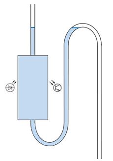
Figure 1 – Structural design of the liquid transparency sensor
The tube for the removal of fluid is inserted into the patient’s urethra at one end and into the reservoir at the other end. It is proposed to use a flask from a dropper to provide the required volume of liquid, and bend the tube in the form shown in Figure 1. New liquid will come from above, falling under the sensor beam, and the old one will gradually come out by filling the left side of the bend. To ensure reliable fastening of the tube, it is proposed to use a special form designed on a 3D printer, in which the sensor will already be fixed. Medical personnel only need to insert the tube into this mold and turn on the power to the device.
The rest of the system works in the same way as in [1]. The implementation of the microprocessor device assumes the use of the ArduinoUno module, based on the ATmega328P processor. Receiving data from the liquid withdrawal control sensor is carried out using a digital port, which is polled by interrupting the level change at the output. Connecting a liquid transparency sensor assumes the use of an ADC module. Empirically, a voltage will be detected, which signals the normal course of the procedure (the liquid is completely transparent). Then, gradually darkening the liquid, a certain scale will be obtained, which can be interpreted as the degree of transparency of the liquid. Based on the obtained boundaries, using the conditions, it is possible to determine the presence of blood in the excreted fluid. In this case, the degree of transparency will signal the urgency of calling a doctor or taking any measures. This parameter is supposed to be transmitted with data on the course of the procedure to the central server.
The hypothesis of the most efficient use of LED color is the use of an infrared emitter, as it is least affected by the degree of illumination in the operating conditions of the device. An infrared emitter was also used in [1] for calculating the volume of the injected solution, and showed good results for use in various rooms, with different degrees of illumination.
References
- Шамков А. С., Чернега В. С. Мир компьютерных технологий: Сборник статей всероссийской студенческой научно-технической конференции, г.Севастополь, 2 – 6 апреля 2018 г/ М-во образования и науки РФ, Севастопольский государственный университет; науч. ред. Е.Н. Мащенко – г. Севастополь: СевГУ, 2018. – с.105-107.
- Теория и практика измерения мутности. – г. Москва, 2020. URL: https://www.ecoinstrument.ru/service/public/teoriya_i_praktika_izmereniya_mutnosti_turbidimetriya_i_nefelometriya2/ (дата обращения: 23.03.2020).
All articles of author «Шамков Андрей Сергеевич»
© If you have found a violation of copyrights please notify us immediately by e-mail.